Today breast cancer is the most usual cancer among women. It mostly affects women between the age of 50 and 70. Its prevalence is rare in women below the age of 30. Every woman during her lifetime will eventually develop some kind of tumor in her breasts. In most of the cases there are benign tumors, such fibroadenomas, pappilomas and fibrocystic breast disease lesions. Nevertheless, 15% of all women will at some point of their life develop breast cancer and according to U.S. statistics, breast cancer is the second most common cause of death among women.
Risk factors for breast cancer include:
• Age (more frequent in older women)
• Family history (relatives with breast cancer)
• Gene mutations (BRCA-1 and BRCA-2 and other oncogenes)
• Beginning of menstruation below the age of 12
• Menopause above the age of 55
• First pregnancy above the age of 35
• Lack of pregnancy and breast feeding
• Existence of atypical breast lesions
• Pre-existing breast cancer
• Previous high dosage radiation in the chest
• Long time and high-dosage intake of oestrogens
• Smoking and obesity
For a successful treatment of the disease early diagnosis is crucial. In those stages we can offer permanent treatment in rates above 95%. This can be achieved by the self-examination of the breasts, regular mammography and ultrasound combined with annual examination by a specialised oncologist surgeon.
Self-examination:
 This must be done almost once in a month, just after the end of menstruation for women of reproductive age. It initially includes the observation of the breasts while standing against the mirror, looking for asymmetry, inlets and bulges. Following, the breasts and the axillaries are self-palpated with small circular movements, using vertical pressure against the thoracic wall.
Radiologic evaluation
 At every age, even during pregnancy if necessary, we can evaluate the breast with ultrasound scan, obtaining valuable diagnostic information. Mammography is indicated above the age of 35, initially as a reference screening and at 45 annually. In some cases more special exams are used such magnetic mammography.
Clinical evaluation:
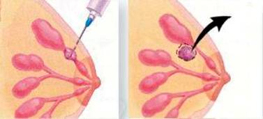
Clinical examination by a specialised oncologist surgeon can combine clinically the palpative and radiologic findings, separating benign from malignant suspected lesions. Every suspected lesion for malignancy is further evaluated with biopsy, either through a small incision or through a fine needle aspiration. In case of aspiration, this can be done even with mammographic or ultrasound guidance.

Surgical treatment
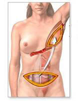
The type of the surgical procedure is defined by the location, the size and the nature of the tumor to be removed. As a general rule, early diagnosed neoplasms rarely require total mastectomy. On the other hand, tumors with delayed diagnosis often require total mastectomy. In that case there are special plastic surgery techniques that can be applied, in order to cover the missing of the breast volume and the skin, creating a new breast.
Crucial for the course of the disease and the long-term survival of the patient is the lymph node dissection. Breast is drained in three main lymphatic groups in the axillary area. The complete excision of all three groups is possible by specialised surgical oncology teams, for the achievement of a radical procedure, which drastically reduces the recurrence rates, while it impressively improves life expectancy. At an early stage, the sentinel lymph node technique can be applied, which is to detect the node that initially drains the tumor. If it is free of metastases, then the axillary dissection can be excluded, simplifying that way the operation.


Post-surgical treatment
After surgery, usually chemotherapy and hormone therapy follows. In case of lymph node metastases, radiotherapy is added. All of the above treatments are well tolerated by the patients, who don’t need to change their daily routine, maintaining their personal, professional and family activity intact.


